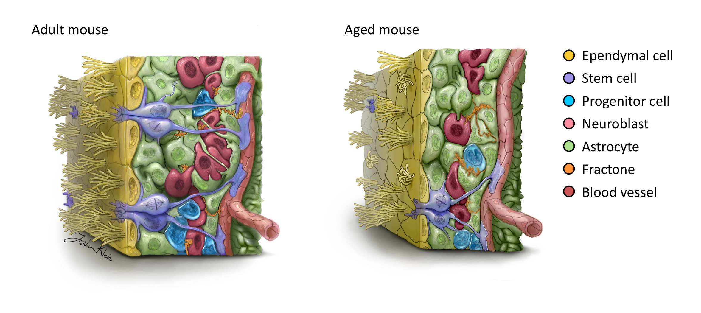
One focus of our research is to characterize the development of the stem cell niche, the ventricular-subventricular zone (V-SVZ), that lines the lateral wall of the lateral ventricles of the brain.
During development, the stem cell niche generates new neurons and supporting glial cells to neighboring brain regions, and ependymal cells to line the ventricle walls. The ependymal lining consists of a single layer of ciliated epithelial cells, which play a role in regulating cerebrospinal fluid (CSF) and interstitial fluid (ISF) homeostasis within the ventricles and the brain, which facilitates CSF circulation and metabolic waste clearance essential to a healthy ventricular system.


Fetal hydrocephalus is a condition characterized by accumulation of CSF, expansion of the lateral ventricles, and associated cognitive defects.
- Alterations in ventricle volume in the developing brain pose a significant problem, as they occur during the period of active stem cell proliferation, neurogenesis, neuroblast migration and ependymal cell formation, ependymogenesis.
- We are investigating how these developmental processes are affected in the hydrocephalic brain.
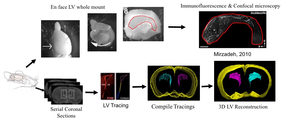
- To characterize brain development in hydrocephalus, we use a combination of immunohistochemistry and ventricle volume analyses to generate 4D spatiotemporal maps of ventricle development and associated stem cell niche dynamics.
- These spatiotemporal maps will provide valuable information to improve diagnostic tools and develop appropriate therapies for hydrocephalic patients.